New equipment: In line with Eye & Retina Surgeons’ commitment to on going investment in the latest and best equipment, we have recently acquired:
Cirrus Optical Coherence Tomography (OCT) from Carl Zeiss: State-of-the-art 3rd Generation Macular & Optic Nerve Scans
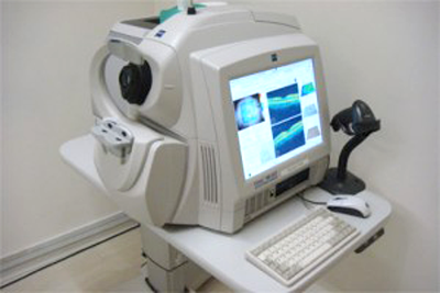 |
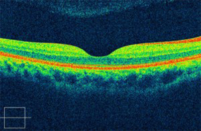
ERS is proud to unveil the latest and most sophisticated technology in OCT (optical coherence tomography) imaging of the macula (centre of the retina) and optic nerve using the Cirrus-HD OCT. Cirrus HD-OCT realizes the famed superior capabilities of ZEISS optics and provides exquisite high definition images and analyses for enhanced clinical confidence.
Beautiful high definition OCT scans and LSO fundus images provide visualization of retinal structure. HD layer maps and thickness maps reveal the critical details of histology and pathology at a glance. The images can be rendered in 3D format.
 |
The Cirrus-HD OCT also boasts enhanced efficiency and speed, reducing time required for a macular scan from 2 minutes to about 30 seconds. In addition, advanced hardware and software facilitate automated alignment and high patient through-put. All these innovations mean increased comfort and convenience for patients.
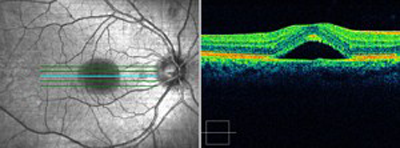 |
Simultaneous capture of the OCT and the LSO fundus image ensures precise registration between the OCT scan and the fundus image.
Repeatability is another important feature of the Cirrus-HD OCT. Automated visit-to-visit registration ensures reproducibility with overlay of the OCT fundus image from the previous scan for verification of scan position. The auto Patient Recall™ assures patient position and instrument setting are repeated from previous visit.
 |
The RNFL thickness map shows the patterns and thickness of the nerve fiber layer. RNFL thickness is displayed in graphic format and compared to normative data
Its accurate repeatability makes it indispensable in the management and monitoring of patients with conditions such as macular degeneration, diabetic retinopathy and glaucoma.
The nidek computerised refractor workstation
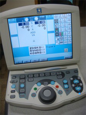
The Nidek Computerized Refractor Workstation is a fully computerized diagnostic equipment to measure a person’s refraction (spectacle prescription). It includes an autorefractor, a lensometer, a phoropter and a chart projector that is fully integrated in form and in function, in a cutting edge platform that incorporates the latest technology available. Using sophisticated computer programs built into the system, it enables the ophthalmologist or optometrist to measure a person’s spectacle prescription and refine it to a high level of accuracy and consistency. The workstation is also ergonomic and patient-friendly to ensure that the measurement process is done safely, efficiently and comfortably for every person.
 |
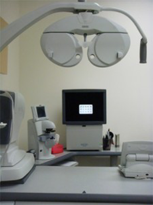 |
The Nidek Autophoropter system for more accurate refraction (measurement of spectacle power)


